ECR 2017 / C-2406
Increased nuchal translucency - Radiological and pathological review of a case series
This poster is published under an open license. Please read the disclaimer for further details.
Congress:
ECR 2017
Poster Number:
C-2406
Type:
Educational Exhibit
Keywords:
Obstetrics (Pregnancy / birth / postnatal period), Forensic / Necropsy studies, Foetal imaging, Ultrasound, Conventional radiography, Intrauterine diagnosis, Foetus, Pathology
Authors:
D. Castelo1, M. Marinho1, F. C. Pires1, S. Florim1, R. Nogueira2, C. Maciel3, F. Valente4; 1Vila Nova de Gaia/PT, 2Braga/PT, 3Porto/PT, 4Gaia/PT
DOI:
10.1594/ecr2017/C-2406
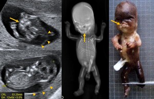
 associated with diffuse subcutaneous thickening and pulmonary edema (long arrow). This exquisite appearance was believed to be secondary to congenital cardiac anomalies, notice an absent a wave at ductus venosus flow spectrum (spaced Doppler complexes). Other findings can be appreciated: "sandal gap" sign (left foot) and calcified/echogenic bowel loops (arrow heads). Flexed limbs suggesting arthrogryposis. References: CGC Genetics and Centro Hospitalar de Vila Nova de Gaia e Espinho")
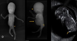
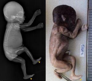
Fig. 6:
Down syndrome case terminated at 15 weeks. Ultrasound, radiographic and...
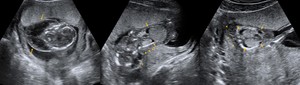
 of a 13 week fetus with Down syndrome. Note de thickened nuchal soft tissues (short arrows) and the absent nasal bones (asterisk). One can also apreciate a right sided club foot deformity (long arrow).")
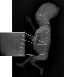
Fig. 7:
Foetal radiography (lateral view) of a 13 week fetus with Down syndrome. Note...
.")
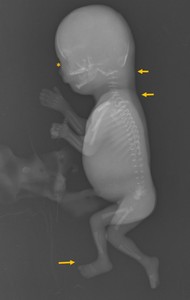
Fig. 8:
A case of Down syndrome, terminated at 15 weeks. Lateral radiography where...
, extending caudally as diffuse subcutaneous thickening (arrow heads). Also notice the bilateral pulmonary edema.")
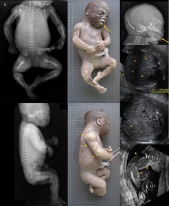
Fig. 11:
Turner syndrome at 12 weeks of pregnancy - note the large cystic hygroma at the...
, omphalocele (long arrows), iliac bone dysplasia (arrow head) and absent nasal bone (short arrow).")
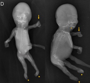
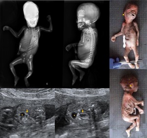
Fig. 9:
Edwards syndrome at 14 weeks. The main anomalies identified were growth ...
, bilateral club feet and clenched hands.")
Fig. 10:
Edwards syndrome - frontal and lateral foetal radiography, aplasia radii of the...
 and cystic hygroma (arrow heads) can be appreciated. References: CGC Genetics and Centro Hospitalar de Vila Nova de Gaia e Espinho")
Fig. 12:
Patau syndrome terminated at 18 weeks - growth retardation, facial cleftings...
, dilated bowel loops were observed (arrows). Normal sized urinary bladder (asterisk). At the fetal radiography 13 ribs can be counted bilaterally. References: CGC Genetics and Centro Hospitalar de Vila Nova de Gaia e Espinho")
Fig. 14:
Cat-Eye syndrome - termination of pregnancy at 24 weeks. Pre-auricular tags and...
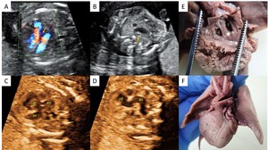
 A - auriculo-ventricular concordance and a large sub-valvular ventricular septum defect (arrow); B - hypoplastic pulmonary artery (arrow); C and D aorta (ao) originating from de right ventricle (vd)(ve - left ventricle); E and F autopsy specimens confirming the ultrasound findings. References: CGC Genetics and Centro Hospitalar de Vila Nova de Gaia e Espinho")
Fig. 16:
Cardiac malformation - foetal ecocardiography (A to D) A - auriculo-ventricular...

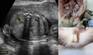
Fig. 15:
Undetermined syndrome - lateral radiographic view and macroscopy photography....
. Dilated urethers (arrow heads), lobulated asymmetric kidneys, micropenis with hypospadias and anal atresia (small arrow) with dilated bowel loops (large arrow) were visualised at autopsy. References: CGC Genetics and Centro Hospitalar de Vila Nova de Gaia e Espinho")
Fig. 17:
Undetermined syndrome 22 weeks - bilateral pyelectasis (ultrasound image -...
; hepatomegaly seen at different levels (asterisks) and macrossomic aspect of the fetus, notice the overdeveloped muscles of the upper limb (arrow heads). References: CGC Genetics and Centro Hospitalar de Vila Nova de Gaia e Espinho")
Fig. 13:
Beckwith-Wiedemann syndrome - large protruding tong (arrows); hepatomegaly seen...
(q32)dn.ish, a rare and not well characterized genetic syndrome. References: CGC Genetics and Centro Hospitalar de Vila Nova de Gaia e Espinho")
Fig. 18:
Turricephaly - notice de elongated head, resulting from premature closure of...
 and diffuse subcutaneous and visceral lymphangiomatosis (note the abdominal distension), confirmed at fetal autopsy.Termination was performed at 16 weeks of pregnancy.")
Fig. 19:
Undetermined syndrome manifesting a large cystic hygroma (arrows) and diffuse...